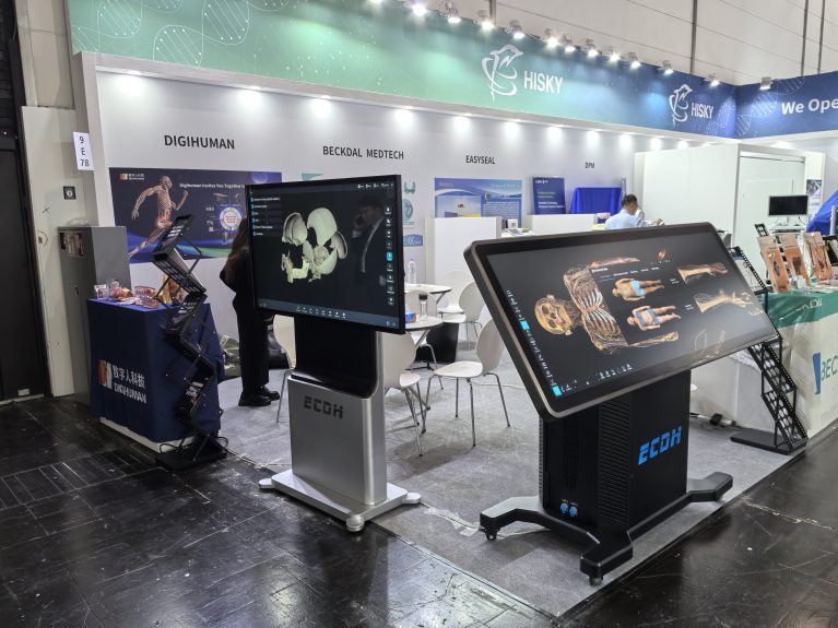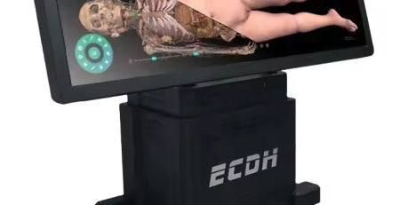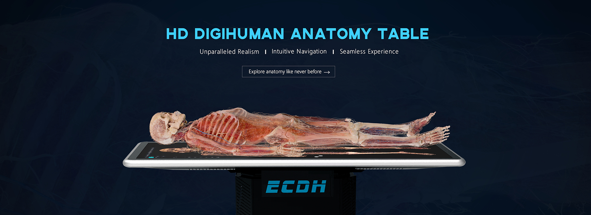
HD Digihuman Virtual Anatomy Table
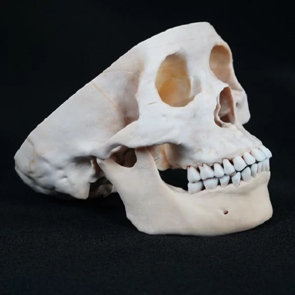
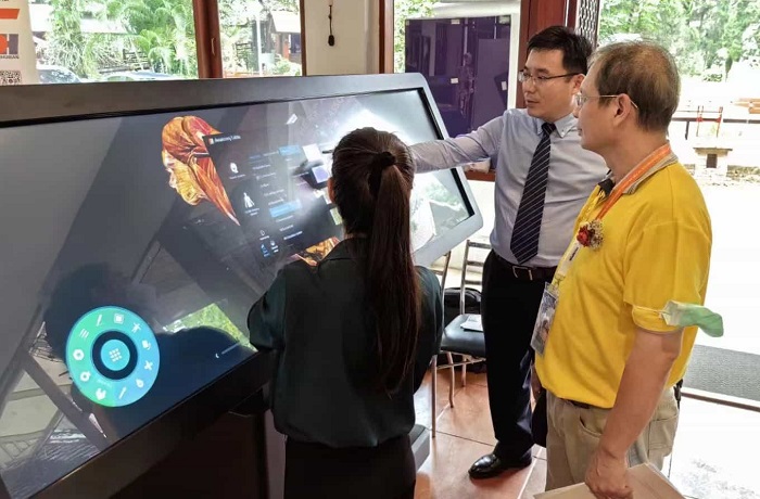
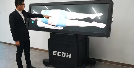
The HD Digihuman Virtual Anatomy Table adopts real human body data reconstruction, simulates the real anatomy process, combine traditional anatomy teaching with technology to provide a idealistic learning solution. The screen can be lifted and tilted to meet different teaching scenarios.
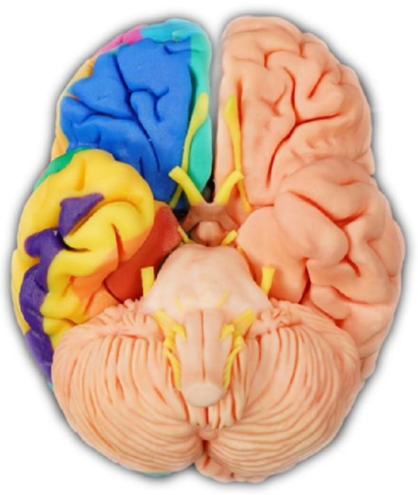
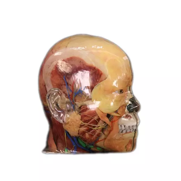
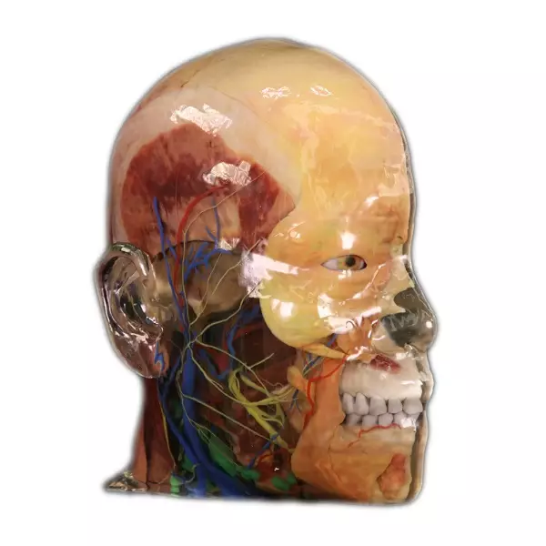
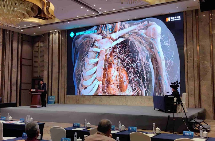
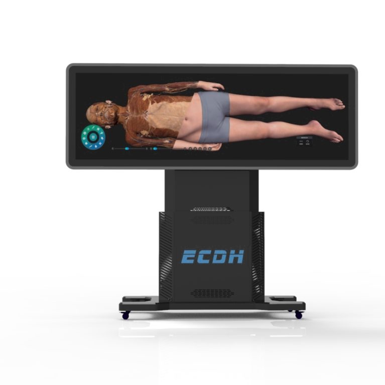
- In this Virtual Autopsy Table’s system, human structures can display in 3D, which can be enlarged.reduced and rotated at any angle.
- The images in Virtual Autopsy Table include CT images and MRI images. The images are sequence images, which can be browsed. Each case contains basic information, main complaint, imaging findings, imaging diagnosis, etc.
- Multiple user accounts can be created, and selected preset can be customized, collection; Set Key images can be set and collected: 3D structures can be Marked.DICOM view history can be reviewed.
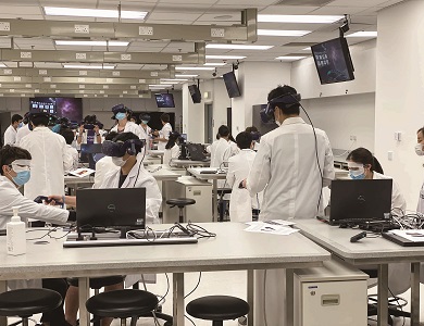
- Each anatomical structure is annotated with description and key structureannotation, and can be hidden, transparent, separately displayed, locked, dyed, multiselected,etc. Perfect for virtual reality anatomy education.
- Hardware:
- Screen:Size:88-inche, Resolution:3840*1080; Brightness: 700cd / ㎡Contrast: 1100:1: Touch mode: infrared touch.
- Computer: I7 above generation 10 /64G DDR4 3200/2T NME SSD/RTX3080 above 10G /in10
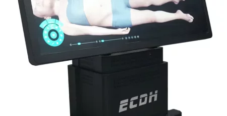
- Screen electric lifting: The 88-inch display can be raised and lowered vertically and can be reversed 90 degrees; The 88-inch display can be reversed90 degrees.
- Our 3d anatomy tables have CE and FCC certifications.