In our pursuit of innovative educational tools, we are excited to present the Digihuman 3D Printing Model-Lumbar Disc Herniation Model. This advanced model is designed to bridge the gap between theoretical knowledge and practical application, specifically addressing the complexities associated with disc herniation. Our commitment to accuracy and detail ensures that healthcare professionals can confidently use this model for surgical planning and improved doctor-patient communication.
Accurate Replication of Anatomical Structures
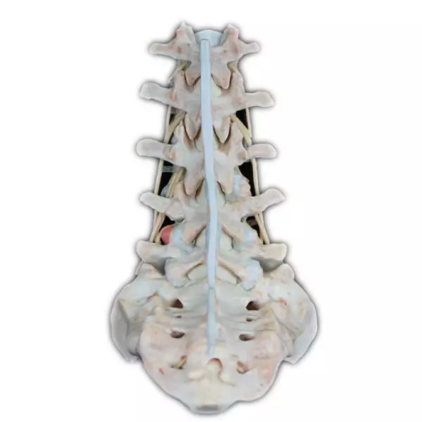
The Digihuman disc herniation model is crafted from a combination of soft and hard materials, which allows it to closely mimic real patient data. By incorporating both normal anatomical structures and various pathological symptoms, this model serves as an invaluable resource for medical professionals. Every aspect of the model has been meticulously developed using high-precision digital human data sets, including original sectional data and refined segmentation data.
With a voxel size of 0.0384mm x 0.0384mm x 0.1mm, the level of detail provided in the lumbar disc herniation model is exceptional. It includes key anatomical features such as bones, muscles, blood vessels, nerves, and ligaments, all accurately represented to reflect real-life human anatomy. This fidelity ensures that users can understand the intricacies associated with disc herniation, facilitating better decision-making during clinical evaluations and surgeries.
Enhanced Surgical Planning and Patient Communication
One of the principal advantages of the Digihuman disc herniation model is its utility in surgical planning. Surgeons can use the model to visualize the specific characteristics of a patient’s condition, making it an essential tool for pre-operative assessments. The realistic representation of disc herniation enables practitioners to devise customized surgical strategies according to individual patient needs.
Moreover, the model enhances communication with patients. By using the disc herniation model, doctors can provide patients with a tangible reference to explain their conditions more effectively. This visual aid fosters a better understanding of the issues at hand, ultimately improving the patient experience and ensuring informed consent for procedures.
Conclusion: A New Era in Medical Education
As we continue to innovate in the field of medical education, the Digihuman 3D Printing Model-Lumbar Disc Herniation Model stands out as a critical asset for healthcare providers. Its precise replication of anatomical structures and diverse pathological presentations makes it an indispensable resource for surgical planning and enhanced doctor-patient interactions.
For healthcare professionals aiming to improve their understanding of disc herniation and its implications, we strongly recommend the Digihuman disc herniation model. By integrating this state-of-the-art model into your practice or educational curriculum, you can significantly elevate the standards of care and improve patient outcomes. Join us in advancing medical education with DIGIHUMAN and discover the potential of 3D printing in healthcare.
The Art and Science of 3D Anatomical Modeling at DIGIHUMAN
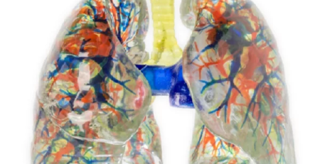
At DIGIHUMAN, we are proud to present our cutting-edge wholesale 3D model of human lungs. Crafted from high-precision digitized human...
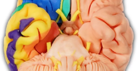
Where to Buy a Brain Model: Your Guide to Digihuman’s 3D Printed Solutions
"Where can I buy a brain model?" People often ask this question. As a leading manufacturer of high-quality anatomical models, we at DIGIHUMAN are...
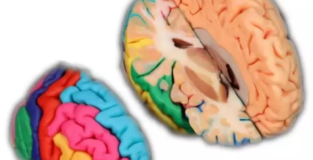
Crafting Realism: The Art and Science of Anatomical Model Creation
At DIGIHUMAN, we take pride in our cutting-edge technology that allows us to create precise anatomical models from digitized human...