As technology continues to evolve, the field of medical education has seen significant advancements. One of the most revolutionary tools in this space is the DIGIHUMAN Anatomy Digital Table. This cutting-edge device is transforming how medical students, educators, and professionals interact with anatomical structures. By providing an immersive and highly interactive learning experience, the anatomy digital table is changing the way the human body is studied.

Revolutionizing Medical Education with the DIGIHUMAN Anatomy Digital Table
The DIGIHUMAN Anatomy Digital Table plays a pivotal role in virtual dissection, allowing students to study human anatomy in high definition without the need for traditional cadavers. Virtual dissection brings numerous benefits to medical education, such as offering endless opportunities for exploration and reducing ethical concerns related to physical dissections.
The table offers high-definition 3D anatomical visualization, enabling users to examine every layer of the body from skin to bones, muscles, and organs. This high level of detail supports a deeper understanding of human anatomy and pathology. Furthermore, the interactive learning features of the DIGIHUMAN Anatomy Digital Table make it an ideal tool for immersive teaching. Medical instructors can utilize the table to display various anatomical structures, engage students with hands-on learning, and create interactive quizzes or exercises to solidify the concepts.
Comprehensive and Detailed Section Library for Medical Training
Another standout feature of the DIGIHUMAN Anatomy Digital Table is its extensive and comprehensive section library. The morphological section provides access to histological and pathological samples, categorized according to tissues and organs. With over 2,000 sections available, users can delve into a vast array of specimens and study them in detail. Whether you’re looking for tissue-specific samples or seeking a particular part of the body, the table allows for easy navigation and exploration.
The DIGIHUMAN Anatomy Digital Table is equipped with tools for efficient search and classification, making it easier for students and researchers to find the exact section they need. The ability to zoom in and analyze tissues at various magnifications—4X, 10X, 20X, 40X—ensures an unparalleled level of detail for medical studies.
Advanced Medical Imaging with DIGIHUMAN
In addition to its anatomical section library, the DIGIHUMAN Anatomy Digital Table integrates powerful medical imaging features. It supports CT and MRI sequence images, allowing for realistic views of internal structures. The table’s high-resolution medical images enable users to study anatomy from multiple perspectives, including sagittal, coronal, and axial views.
A key feature of the DIGIHUMAN Anatomy Digital Table is its support for DICOM (Digital Imaging and Communications in Medicine) information, which enables users to view and adjust images based on their specific requirements. Moreover, the ability to reconstruct 3D images from the CT and MRI scans offers a deeper, more comprehensive understanding of the human body. This feature allows students and professionals to observe organs and structures from various angles and even cut through the 3D models for more focused studies.
Conclusion
The DIGIHUMAN Anatomy Digital Table is more than just a tool; it’s an integral part of modern medical education. By offering high-definition anatomical visualization, a comprehensive section library, and advanced medical imaging features, it creates an unparalleled learning experience. Whether you’re a student, educator, or medical professional, the anatomy digital table from DIGIHUMAN is revolutionizing the way we learn and understand the human body.
The Art and Science of 3D Anatomical Modeling at DIGIHUMAN
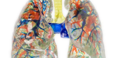
At DIGIHUMAN, we are proud to present our cutting-edge wholesale 3D model of human lungs. Crafted from high-precision digitized human...
Where to Buy a Brain Model: Your Guide to Digihuman’s 3D Printed Solutions
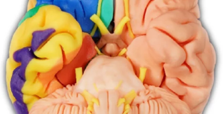
"Where can I buy a brain model?" People often ask this question. As a leading manufacturer of high-quality anatomical models, we at DIGIHUMAN are...
Crafting Realism: The Art and Science of Anatomical Model Creation
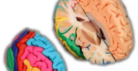
At DIGIHUMAN, we take pride in our cutting-edge technology that allows us to create precise anatomical models from digitized human...