At DIGIHUMAN, we are dedicated to advancing medical education through innovative technologies. Our latest offering, the Digihuman 3D Printing Model-Lung Segment, represents a significant breakthrough in the way anatomical structures are visualized and understood. By constructing a multi-structured 3D digital anatomical model derived from high-precision digitized human body data, we provide educators and learners with an unparalleled tool for exploring human anatomy.
A Highly Detailed Lung Model
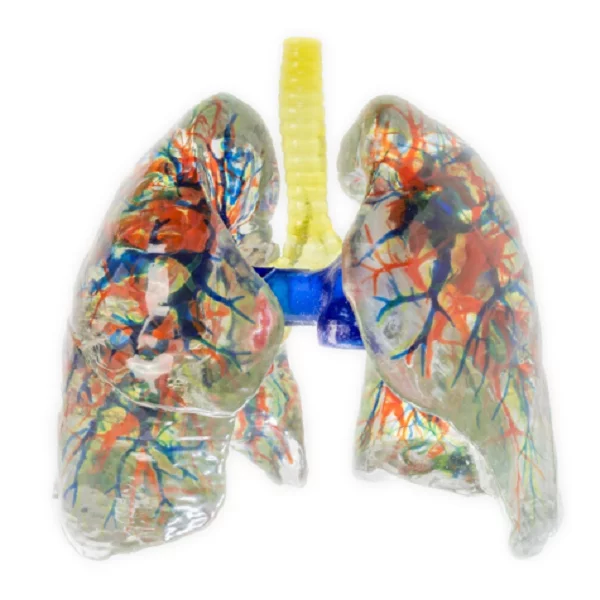
The Digihuman 3D Printing Model-Lung Segment is crafted using a full-color, multi-material 3D printer, utilizing environmentally friendly resin materials. This allows us to create a lung model that achieves a realistic 1:1 high simulation of physical anatomical structures. Each component of the lung model is meticulously designed to ensure accuracy and detail, making it an essential resource for educational institutions and healthcare professionals alike.
One of the key features of our lung model is its detachable combined design, composed of transparent wrapping material. This model includes crucial structures such as the bronchial tree, pulmonary artery, and pulmonary vein, allowing for a comprehensive understanding of the respiratory system. The use of color-coding—blue for the pulmonary artery, red for the pulmonary vein, and various colors for the bronchus segments—enhances visual learning and helps students easily identify different parts of the lung.
Comprehensive Anatomy Learning Tool
Education in anatomy relies heavily on practical and visual aids. The lung model serves as an invaluable resource for both instructors and students. By segmenting the lung into eight segments for the left lung and ten for the right lung, we have created a split and combined lung segment model that facilitates hands-on exploration. This structure allows students to engage with the lung model interactively, enabling deeper learning as they can manipulate and examine each part of the model in detail.
This lung model not only assists in demonstrating the functional anatomy of the lungs but also fosters a better understanding of clinical applications. Healthcare professionals can utilize this model for training purposes, enhancing their ability to visualize and comprehend complex procedures involving the respiratory system.
Commitment to Advancing Medical Education
As we strive to improve the tools available for anatomical education, the Digihuman 3D Printing Model-Lung Segment stands out as a sophisticated teaching aid. Our commitment to providing high-quality educational resources is unwavering, and we believe that this lung model will significantly enhance learning experiences in anatomy, medicine, and healthcare.
In conclusion, we encourage educators and medical institutions to integrate the Digihuman 3D Printing Model-Lung Segment into their curricula. By utilizing our innovative lung model, you will empower students and professionals with superior knowledge and understanding of human anatomy, ultimately contributing to better healthcare outcomes. Choose DIGIHUMAN as your partner in advancing anatomical education through state-of-the-art resources.
Navigating the Challenge: Is Anatomy and Physiology Hard?
As we embark on our journey into the fields of healthcare and biology, many students grapple with a common concern:...

Sustainable Solutions in Education: The Digihuman Hand Skeleton Model with Muscles
We at DIGIHUMAN are committed to pushing the boundaries of 3D printing technology to enhance educational experiences in anatomy and medical...
Realistic Hand Muscles 3D Model: Transforming Learning Experiences
At DIGIHUMAN, we are committed to advancing the field of anatomical education through innovative technology. Our offering, the Digihuman 3D Printing...