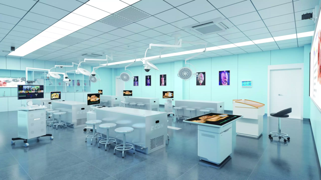
Virtual anatomy tables are now becoming an essential tool for medical training, with leading universities such as Harvard and Johns Hopkins already integrating them into their programs. These tools give students interactive access to human anatomy in ways traditional cadavers cannot, making them highly useful in modern classrooms.
If you want to invest in the best virtual anatomy table, how should you choose? It’s a priority for most medical universities and training institutions, since the right model directly influences teaching efficiency and the quality of student learning outcomes.
Without further ado, keep reading.
What’s the Best Virtual Anatomy Table for Medical Education?
Virtual dissection tables on the market vary more or less in terms of features. Among the many functions, the most critical ones are:
1. Accurate Anatomical Models
The foundation of any virtual anatomy table lies in the accuracy of its models.
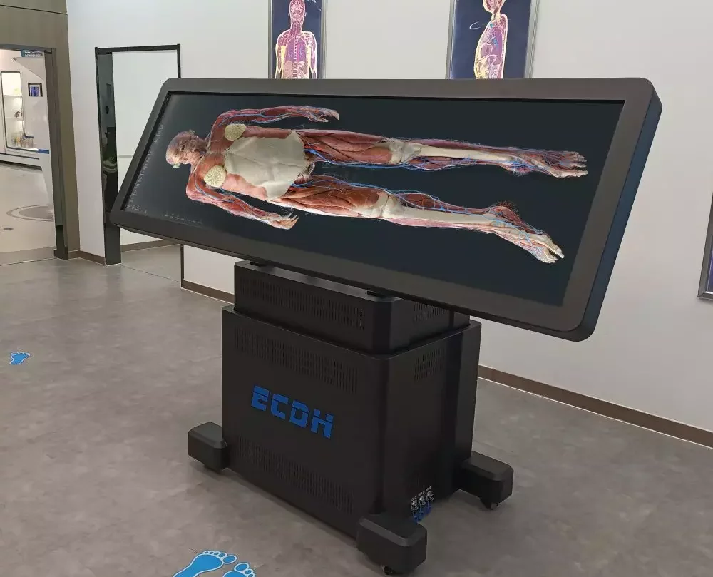
- Real human data:The virtual anatomy table must be constructed based on real human tomographic imaging data to ensure accurate structural restoration and clear layering.
- Model precision: Models should be precise enough to showbones, muscles, vessels, and organs exactly. The finer the detail, the greater the benefit for learners who need to recognize subtle structures that are critical in surgery or diagnosis. For example, the Digihuman Virtual Anatomy Table can achieve an accuracy of 0.1mm to 1mm, and can display over 6,000 anatomical structures in 3D form.
- Observation of fine structures:In addition to macroscopic anatomical structures, the observation of fine structures is also one of the important criteria, such as digital histological sections, thereby meeting multi-level teaching needs from systemic anatomy to detailed study.
2. Teaching Resources
- Multidisciplinary applicability:The best virtual anatomy tables support teaching across multiple disciplines, such as anatomy, pathology, embryology, and animal anatomy, and offer abundant clinical cases. Moreover, by equipping corresponding CT and MRI images alongside tomographic specimen images, it can help students better understand the relationship between radiology and anatomy.
- Test question banks:Another valuable feature to look for is the integration of test question The practice content includes theoretical exercises and specimen exercises, helping teachers better evaluate students’ learning outcomes and effectively extend classroom interaction and self-directed learning.
3. Interaction Methods
How students interact with the digital content is just as important as the accuracy of the models themselves.
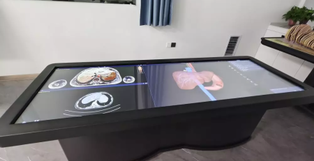
- Touch control:A well-designed digital dissection table should allow users to manipulate structures through simple touch controls, such as zooming in on small nerves, rotating organs for different viewpoints, or performing layered dissections to study internal details step by step. This deepens understanding by exploring spatial relationships.
- Ergonomic designs:Height adjustment and tilting options make the table usable in both lecture halls and smaller lab environments, ensuring comfort for both standing demonstrations and seated study.
- AR tech: Compatible with AR technology, it will deliver an immersive 3D medical training experience. Students can perform virtual dissections through AR devices, resulting in more durable knowledge retention.
4. User-friendliness
Finally, the ease of operation for any device cannot be overemphasized, as it directly impacts the learning time and willingness to use among both teachers and students.
Features such as clear menus, smooth navigation, and responsive performance make the system practical and appealing for daily use.
The best digital anatomy tables also include clear manuals that help students resolve control issues quickly and navigate the system with confidence. A design that minimizes learning curves ensures adoption across all levels of study.
Get Your Optimal Virtual Anatomy Tables
Ethical, preservation, and maintenance issues associated with human cadavers have prompted many universities and institutions to turn to virtual dissection tables. In this regard, Digihuman is an expert virtual dissection table manufacturer, giving medical students and professionals an immersive and interactive learning experience. Features include:
- Ultra-high-definition human tomographic image data(0.1mm) to show bones, muscles, vessels, and nerves, while also offering real dissection videos and animations that demonstrate procedures in detail.
- More than 1,700 CT and MRI scans are seamlessly integratedwith tomographic specimen images.
- Includes test question resources to enhance knowledge consolidation and retention.
- Supportszooming, rotating, and layered virtual dissections with touch controls.
- Compatible with AR devices to create immersive learning
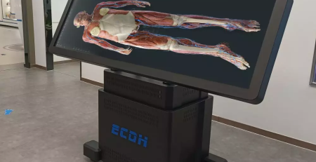
- The screen can be liftedand tilted to accommodate different teaching scenarios.
The system covers systemic anatomy, regional anatomy, embryology, animal dissection, tomographic images, clinical cases, and more, enabling both teachers and students to accomplish learning and operations on a single device.
Wrapping-Up
Selecting the right virtual anatomy lab table is essential for training future healthcare professionals in an effective way. By combining accurate models, interactive tools, and ease-of-use functions, Digihuman delivers a platform that strengthens both teaching and learning practices. Contact us now to learn more information!

3D Printed Anatomical Model Explained: Concept, Applications, and Advantages
Advances in 3D printing and digital modeling are reshaping many fields. One of the most visible changes can be seen...
Innovative Applications of Virtual Dissection Tables
The rise of the virtual dissection table has transformed how anatomy is taught in medical institutions. Traditional dissection has long...
A Comprehensive Guide to Virtual Dissection Table
The anatomy lab has long been an essential venue for cultivating medical students' practical skills. However, it also faces limitations...